Cats are renowned for their acrobatic prowess and uncanny ability to always land on their feet. The spine and tail of your cat are particularly helpful in this regard. The viral debates of buttered toast vs. cat’s feet humorously test physics with feline dexterity, asserting that cats seem to defy the laws of nature. But what anatomical features of the spine allow cats to perform such feats of agility? This article will explore the sophisticated structure of cats’ vertebral column and tail to uncover just how they achieve their remarkable balance and flexibility.
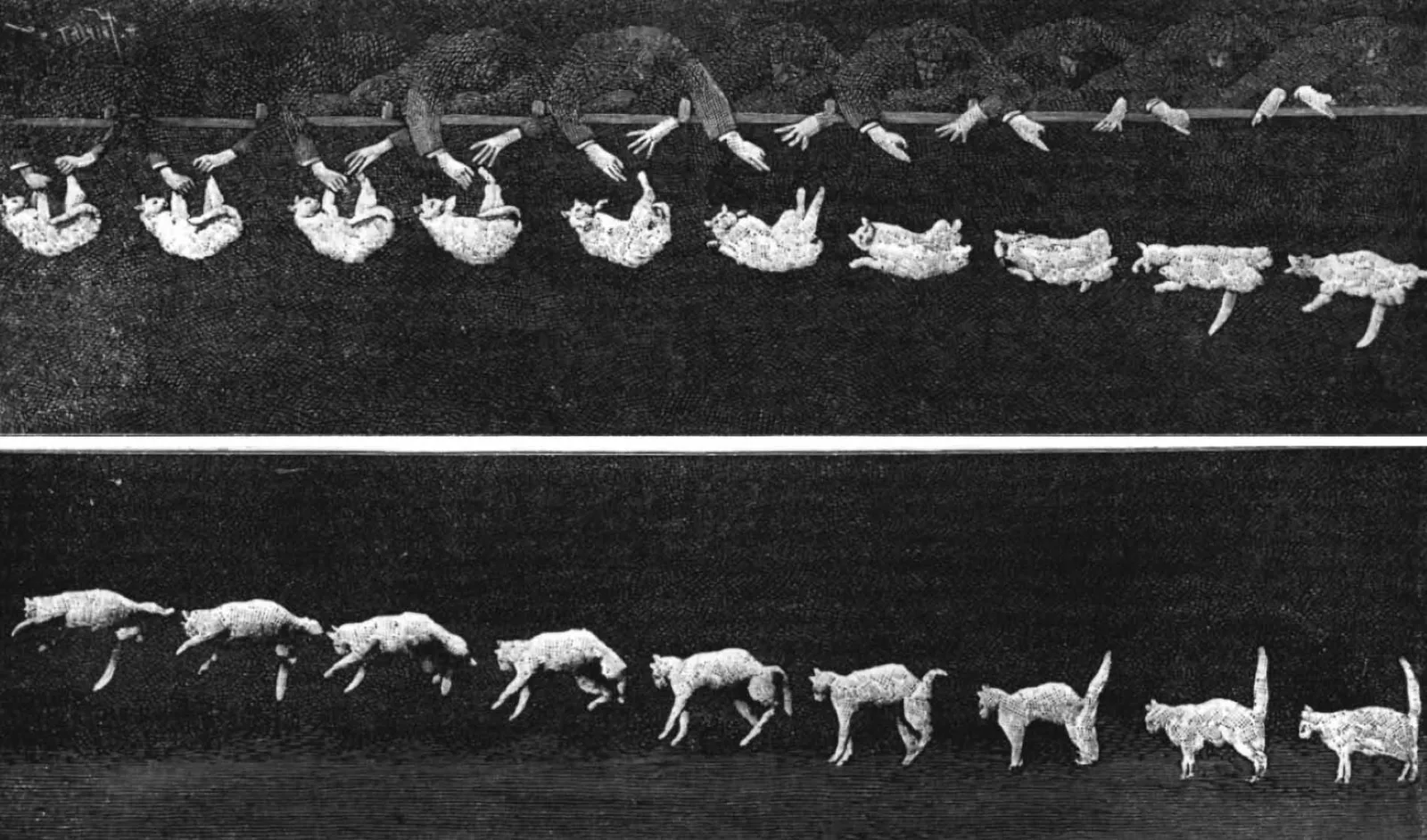
Vertebral column of cats
Cats, like all mammals, have a vertebral column that is segmented into different regions: cervical, thoracic, lumbar, sacral, and caudal. Each segment plays a crucial role in the overall functionality and agility of felines.
What’s special about a cat’s spine?
The cat’s spine consists of more vertebrae than that of a human. Also, unlike humans, whose spinal structure is designed to support an upright posture, the feline spine is optimized for horizontal agility. Cats’ lumbar and thoracic vertebrae are more loosely connected, which provides an enhanced range of motion ideal for sprinting and leaping. Even though cats are physically able to walk on their hind legs for a short amount of time, their preferred movement is on all fours.
Functions of the cat spine
- Support: The spine provides a foundational structure for the body.
- Protection: It shields the spinal cord, a crucial part of the central nervous system.
- Flexibility: The spine’s adaptability is key to a cat’s agility, allowing for high degrees of twisting and bending essential for their dynamic movements.
Spinal regions and how many bones a cat’s spine has
The spine comprises 48 to 53 bones, accounting for about one fifth of all the bones in a cat’s skeleton. Let’s examine its sections more closely.
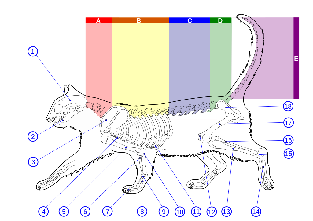
- A – Cervical spine (neck)
This region consists of 7 bones, which allow for a wide range of head movements. This flexibility is critical for cats as it enhances their ability to scan their environment and aids in their predatory skills. - B – Thoracic spine (upper back)
Comprising 13 vertebrae, this area is rigidly connected to the ribs, which aids in stability and protects vital organs. The limited flexibility in this region is compensated by the mobility of the cervical and lumbar regions. - C – Lumbar spine (lower back)
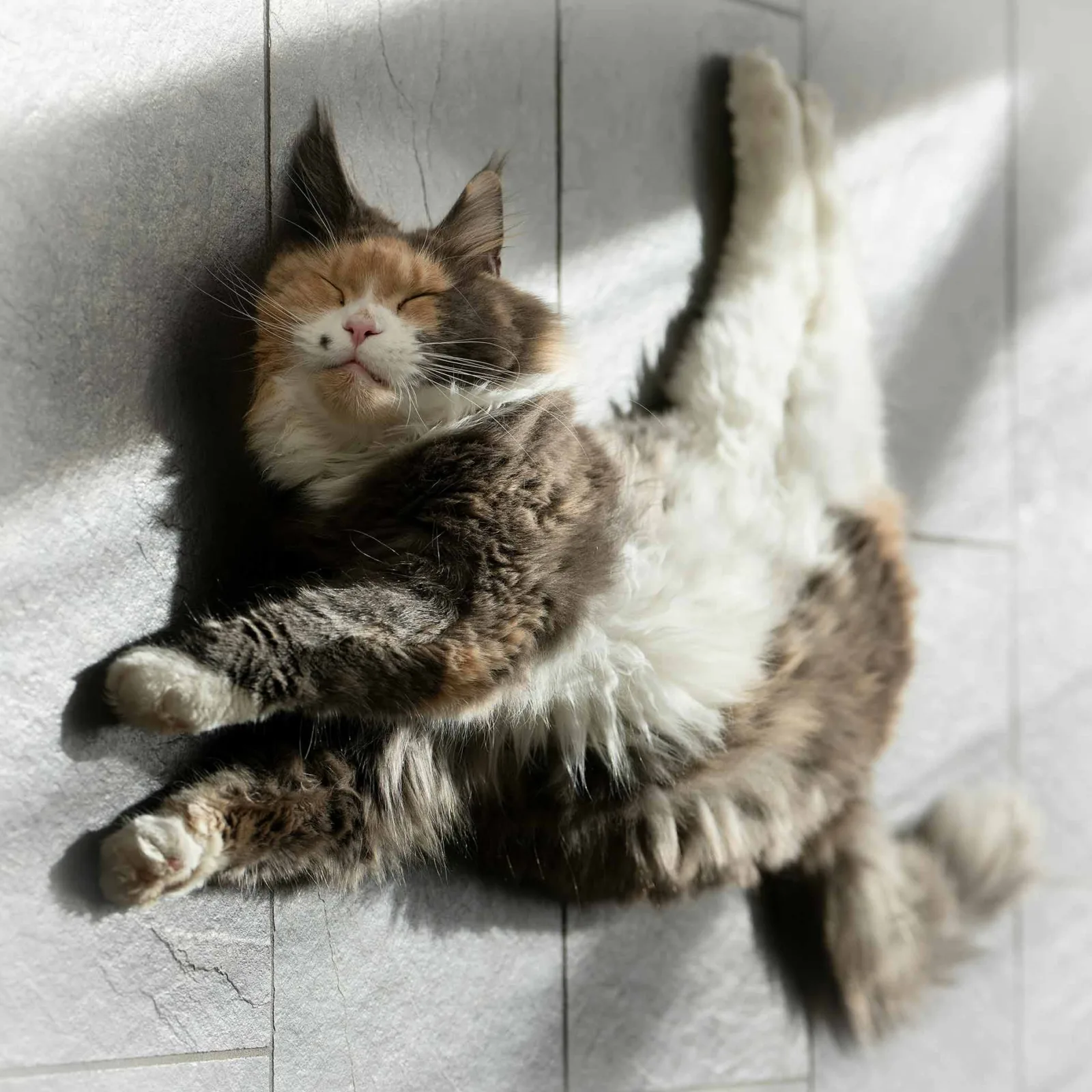
The 7 lumbar vertebrae are key to the powerful muscle attachments that facilitate explosive leaps and fast running. This region’s structure supports high-speed motions and quick directional changes. The possible range a cat can twist is almost 180°!1 - D – Sacral region
The sacral spine’s integration with the pelvis affects the hind limbs and plays a significant role in maintaining stability while moving. There are 3 sacreal vertebra bones. - E – Caudal vertebrae (tail)
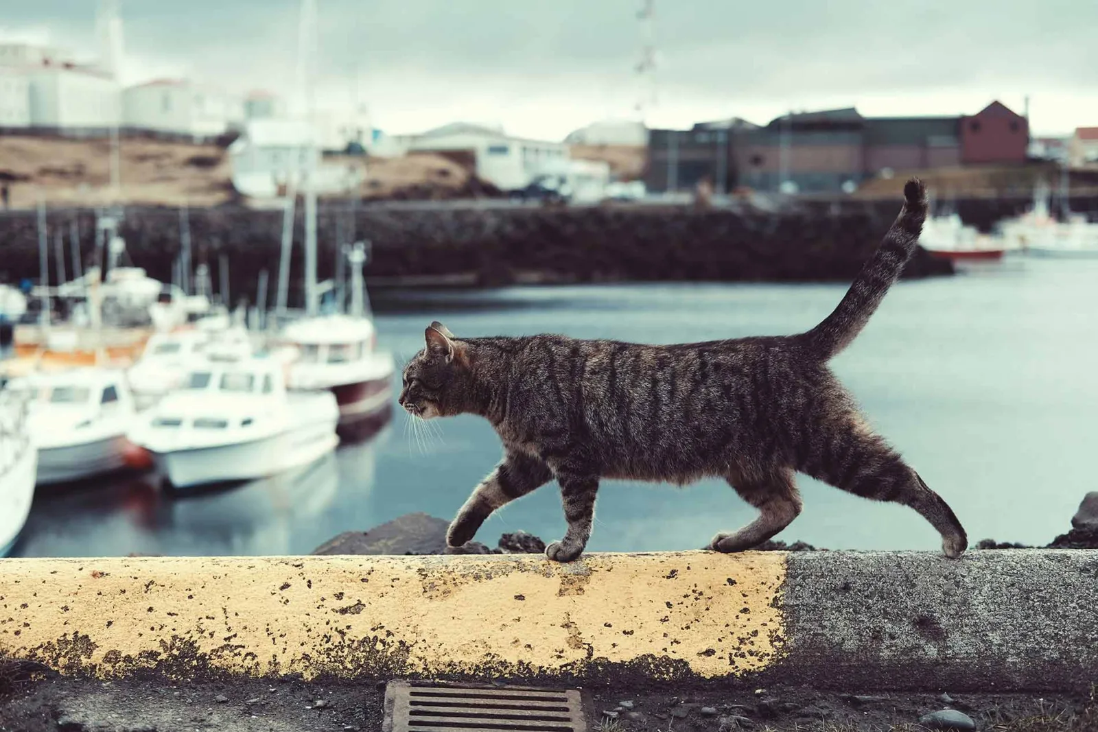
Cats can have between 18 and 23 caudal vertebrae, which significantly contribute to their balance and communicative abilities. The tail acts as a counterbalance during tightrope walks and aids in making sharp turns and quick direction changes.
The cat tail in detail
Anatomy of the cat tail and its functions
Other than bones, the tail consists of caudal vertebrae, joints, muscles, and nerves, intricately designed to support finely tuned movements. This structure is pivotal in maintaining equilibrium, particularly during aerial turns and while navigating narrow spaces. The further down you go, the smaller the bones become, which explains why the tip of a cat’s tail is especially flexible.
Joints, discs and muscles in a cat’s tail
Joints: The caudal vertebrae in a cat’s tail are connected by various types of joints that collectively enhance its flexibility and functional versatility. Synovial joints, which are the most prevalent throughout the tail, facilitate fluid and extensive movements essential for the tail’s agility. Cartilaginous joints, interspersed among the caudal vertebrae, offer slight flexibility and act as cushions, which are crucial for absorbing impacts during dynamic actions and fine-tuning the tail’s balance.
Muscles: Main groups involved are the sacrococcygeal and sacrocaudal muscles. These muscles allow the tail of a cat to move up and down or sway side to side, helping cats maintain their agility and express their emotions. Additionally, the caudofemoralis muscle, which connects the tail to the hind legs, aids in propelling the cat forward by pulling the thigh backwards, thus playing a crucial role in the cat’s ability to run or jump efficiently.
Intervertebral discs: These are located between the vertebrae throughout the spinal column, including the tail (caudal) vertebrae in animals that have tails. These discs are composed of a tough, fibrous outer layer called the annulus fibrosus and a soft, gelatinous center called the nucleus pulposus.The primary function of intervertebral discs is to act as shock absorbers for the spine. They allow for flexibility and movement in the spine while also cushioning the vertebrae against impacts that occur during normal activities and behaviors like jumping, running, and landing.
Nerves in a cat’s tail and how it aids in communication
Nerves: The caudal spinal nerves (coccygeal nerves) emerge from the spinal cord and extend into the tail. They are responsible for sensory innervation and movement control. Basically, the nerves give the directions to the muscles to enable controlled movements. They also make the tail sensitive to touch, temperature, and pain. When receiving a painful stimulus, they react with reflexes, such as twitching away.
Communication:
- Scent communication: The tail contains scent glands which produce pheromones. Cats use these glands to mark territory by leaving scent trails on surfaces.
- Visual and tactile signals: Cats also use their tails to convey a wide range of emotions and intentions through distinct movements. For example, a raised tail often indicates happiness or confidence.
For a comprehensive guide on interpreting your cat’s tail movements and understanding their communication cues, refer to our detailed article, How to Understand Your Cat. It includes an extensive list of tail movements and explains how to interpret them accurately.


